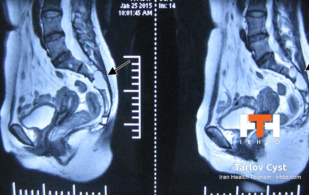
A spinal tumor is an abnormal mass of tissue within or surrounding the cord and/or spinal column. These cells grow and multiply uncontrollably, seemingly unchecked by the mechanisms that control normal cells. Spinal tumors can be benign (non-cancerous) or malignant (cancerous). Primary tumors originate in the spine or spinal cord, and metastatic or secondary tumors result from cancer spreading from another site to the spine.
Spinal tumors may be referred to by the region of the spine in which they occur. These basic areas are cervical, thoracic, lumbar and sacrum. Additionally, they also are classified by their location in the spine into three major groups: intradural-extramedullary, intramedullary and extradural.
Intradural-extramedullary. The most common of these types of tumors develop in the spinal cord’s arachnoid membrane (meningiomas), in the nerve roots that extend out from the spinal cord (schwannomas and neurofibromas) or at the spinal cord base (filum terminale ependymomas). Although meningiomas are often benign, they can be difficult to remove and may recur. Nerve root tumors are also generally benign, although neurofibromas may become malignant over time. Ependymomas at the end of the spinal cord can be large, and the delicate nature of fine neural structures in that area may complicate treatment.
Intramedullary: These tumors grow inside the spinal cord, most frequently occurring in the cervical (neck) region. They typically derive from glial or ependymal cells that are found throughout the interstitium of the spinal cord. Astrocytomas and ependymomas are the two most common types. They are often benign, but can be difficult to remove. Intramedullary lipomas are rare congenital tumors most commonly located in the thoracic spinal cord.
Extradural: These lesions are typically attributed to metastatic cancer or schwannomas derived from the cells covering the nerve roots. Occasionally, an extradural tumor extends through the intervertebral foramina, lying partially within and partially outside of the spinal canal.
Metastatic Spinal Tumors. The spinal column is the most common site for bone metastasis. Estimates indicate that at least 30 percent and as high as 70 percent of patients with cancer will experience spread of cancer to their spine.
Common primary cancers that spread to the spine are lung, breast and prostate. Lung cancer is the most common cancer to metastasize to the bone in men, and breast cancer is the most common in women. Other cancers that spread to the spine include multiple myeloma, lymphoma, melanoma and sarcoma, as well as cancers of the gastrointestinal tract, kidney and thyroid. Prompt diagnosis and identification of the primary malignancy is crucial to overall treatment. Numerous factors can affect outcome, including the nature of the primary cancer, the number of lesions, the presence of distant non-skeletal metastases and the presence and/or severity of spinal-cord compression.
Pediatric spinal tumors. Primary spinal tumors are rare in children and are challenging to treat. Unlike adults, children have not achieved complete skeletal growth, which doctors must take into account when considering treatment. Other factors to consider are spinal stability, surgical versus nonsurgical interventions and preservation of neurological function.
Causes [h4]
The cause of most primary spinal tumors is unknown. Some of them may be attributed to exposure to cancer-causing agents. Spinal cord lymphomas, which are cancers that affect lymphocytes (a type of immune cell), are more common in people with compromised immune systems. There appears to be a higher incidence of spinal tumors in particular
families, so there is most likely a genetic component.
In a small number of cases, primary tumors may result from presence of these two genetic diseases:
Neurofibromatosis 2: In this hereditary disorder, benign tumors may develop in the arachnoid layer of the spinal cord or in the supporting glial cells. However, the more common tumors associated with this disorder affect the nerves related to hearing and can inevitably lead to loss of hearing in one or both ears.
Von Hippel-Lindau disease: This rare, multi-system disorder is associated with benign blood vessel tumors (hemangioblastomas) in the brain, retina and spinal cord, and with other types of tumors in the kidneys or adrenal glands.
Treatment Decisions [h4]
Treatment decision-making is often multidisciplinary, incorporating the expertise of spinal surgeons, medical oncologists, radiation oncologists and other medical specialists. The selection of treatments including both surgical and non-surgical is therefore made keeping in mind the various aspects of the patient’s overall health and goals of care.
Nonsurgical Treatment. Nonsurgical treatment options include observation, chemotherapy and radiation therapy. Tumors that are asymptomatic or mildly symptomatic and do not appear to be changing or progressing may be observed and monitored with regular MRIs. Some tumors respond well to chemotherapy and others to radiation therapy. However, there are specific types of metastatic tumors that are inherently radioresistant (i.e. gastrointestinal tract and kidney): in those cases, surgery may be the only viable treatment option.
Surgery. Indications for surgery vary depending on the type of tumor. Primary spinal tumors may be removed through complete en bloc resection for a possible cure. In patients with metastatic tumors, treatment is primarily palliative, with the goal of restoring or preserving neurological function, stabilizing the spine and alleviating pain. Generally, surgery is only considered as an option for patients with metastases when they are expected to live 12 weeks or longer, and the tumor is resistant to radiation or chemotherapy. Indications for surgery include intractable pain, spinal-cord compression and the need for stabilization of impending pathological fractures.
For cases in which surgical resection is possible, preoperative embolization may be used to enable an easier resection. This procedure involves the insertion of a catheter or tube through an artery in the groin. The catheter is guided up through the blood vessels to the site of the tumor, where it delivers a glue-like liquid embolic agent that blocks the vessels that feed the tumor. When the blood vessels that feed the tumor are blocked off, bleeding can often be controlled better during surgery, helping to decrease surgical risks.
The posterior (back) approach allows for the identification of the dura and exposure of the nerve roots. Multiple levels can be decompressed, and multilevel segmental fixation can be performed. The anterior (front) approach is excellent for tumors in the front of the spine and effectively reconstructing defects caused by removal of the vertebral bodies. This approach also allows placement of short-segment fixation devices. Thoracic and lumbar spinal tumors that affect both the anterior and posterior vertebral columns can be a challenge to resect completely. Not infrequently, a posterior (back) approach followed by a separately staged anterior (front) approach has been utilized surgically to treat these complex lesions.


