

Ophthalmology – A wide variety of disorders can affect the eye and interfere with vision.
Structure of the eye
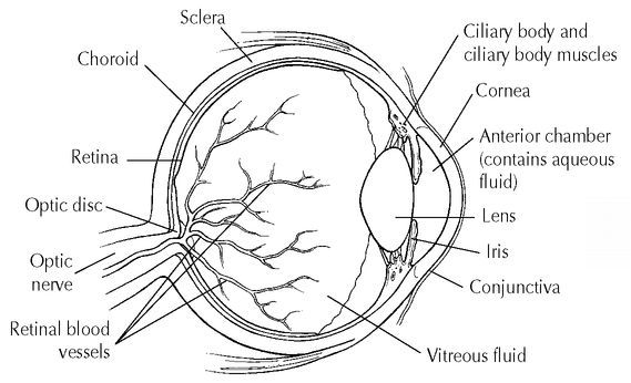
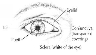
The eye is a complex and delicate structure. Each eyeball is a sphere about 1 inch in diameter, covered by three layers of tissue. The tough, white, outer layer, called the sclera, maintains the shape and size of the eyeball. Covering the sclera at the front of the eye is a transparent mucous membrane called the conjunctiva, which also lines the inside of the eyelids. At the front of the sclera is a transparent, dome-shaped, protective covering called the cornea.
The middle layer of tissue, which lies beneath the sclera, is called the choroid. The choroid contains blood vessels that supply the tissues of the eye with oxygen and nutrients. Toward the front of the eye, the choroid forms a circular ring with muscles called the ciliary body. Attached to the front of the ciliary body is the colored part of the eye, a circular curtain containing muscle fibers, called the iris. In the center of the iris is an opening called the pupil, through which light enters the eye. The dilating (widening) and constricting (narrowing) of the pupil, which are controlled by the muscle fibers of the iris, regulate the amount of light that enters the eye.
Directly behind the iris and pupil is a transparent, elastic structure called the lens, which is attached to the ciliary body. Contraction of the ciliary body muscles changes the shape of the lens and enables the eye to focus. The space between the cornea and the lens is filled with a clear, watery liquid called aqueous fluid. The space behind the lens is filled with a substance called vitreous fluid, which makes up the mass of the inside of the eyeball.
Vision
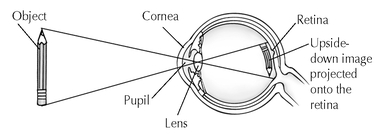
When you look at an object, light rays from the object pass through the cornea, pupil, and lens of the eye, which project an upside-down image of the object onto the retina. The retina converts the image into nerve impulses and transmits the impulses to your brain via the optic nerve. Your brain interprets the information it receives from the retina, and you see the object right side up.
The inner layer of tissue, called the retina, lines the back of the inside of the eye. The retina includes a layer of light-sensitive nerve cells called rods and cones. The rods are very sensitive to light intensity and enable you to see in dim light. The cones detect color and detail. There are 125 million rods and 7 million cones in each eye. When you look at an object, light rays from the object pass through the cornea, pupil, and lens, and form an upside-down image of the object on the retina. The rods and cones transform the sensations of color, form, and light intensity that they receive into nerve impulses, which the retina then transmits along retinal nerve fibers to the optic nerve, a stalklike collection of nerves that connects the eye to the brain. The vision centers in the brain interpret nerve impulses received from each eye and integrate them into the single, right-side-up, three-dimensional image that you see.
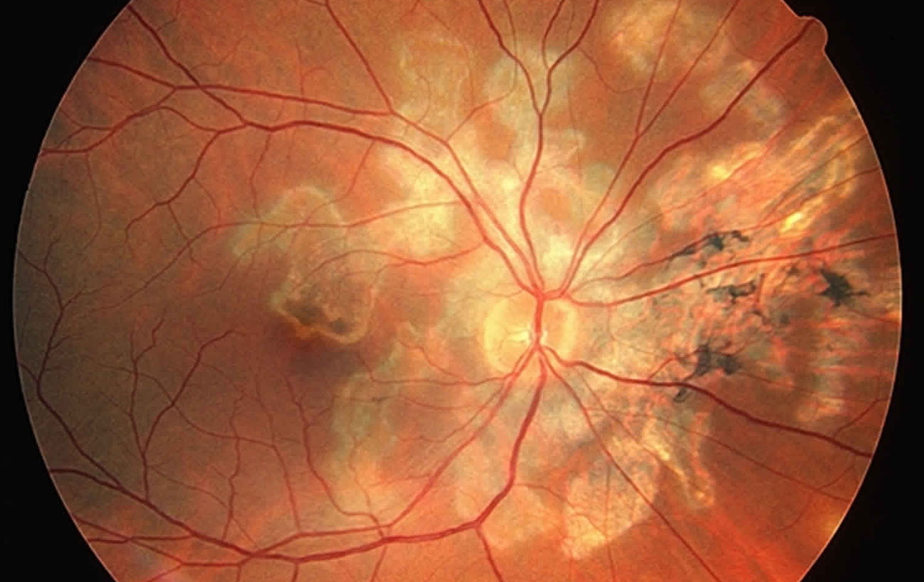
Retinal Artery Occlusion
The retina (the light-sensitive membrane lining the back of the eye) receives its blood supply…
Choroiditis
Choroiditis is inflammation of the choroid, the layer of blood vessels beneath the retina (the…
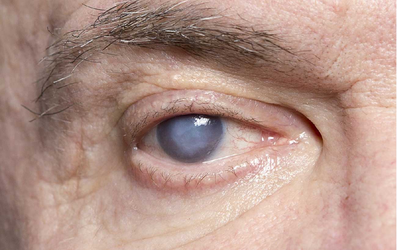
Cataracts
A cataract is clouding of the lens of the eye. As protein fibers in the…

Glaucoma
Glaucoma Glaucoma is a group of eye diseases in which damage to the optic nerve…

Color Vision Deficiency
Color vision deficiency (also called color blindness) is a vision disorder in which a person…
Retinal Vein Occlusion
The central retinal vein carries oxygen-depleted blood away from the retina (the light-sensitive membrane lining…
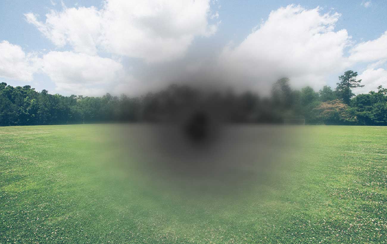
Macular degeneration
Macular degeneration is a disease characterized by irreversible deterioration of the light-sensitive cells of the…

Diabetic Retinopathy
Diabetic retinopathy is an eye disorder in which changes in the blood vessels of the…
Secondary Tumors
Cancer cells can spread through the bloodstream or the lymphatic system from a tumor in…

Optic Neuritis
Optic neuritis is inflammation of the optic nerve, which disrupts the flow of signals from…
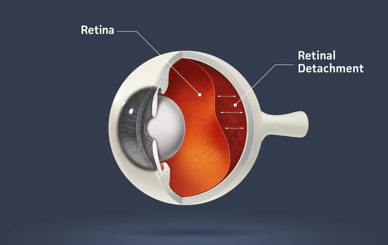
Retinal Detachment
Retinal detachment occurs when the retina—the light-sensitive membrane that lines the inside of the back…
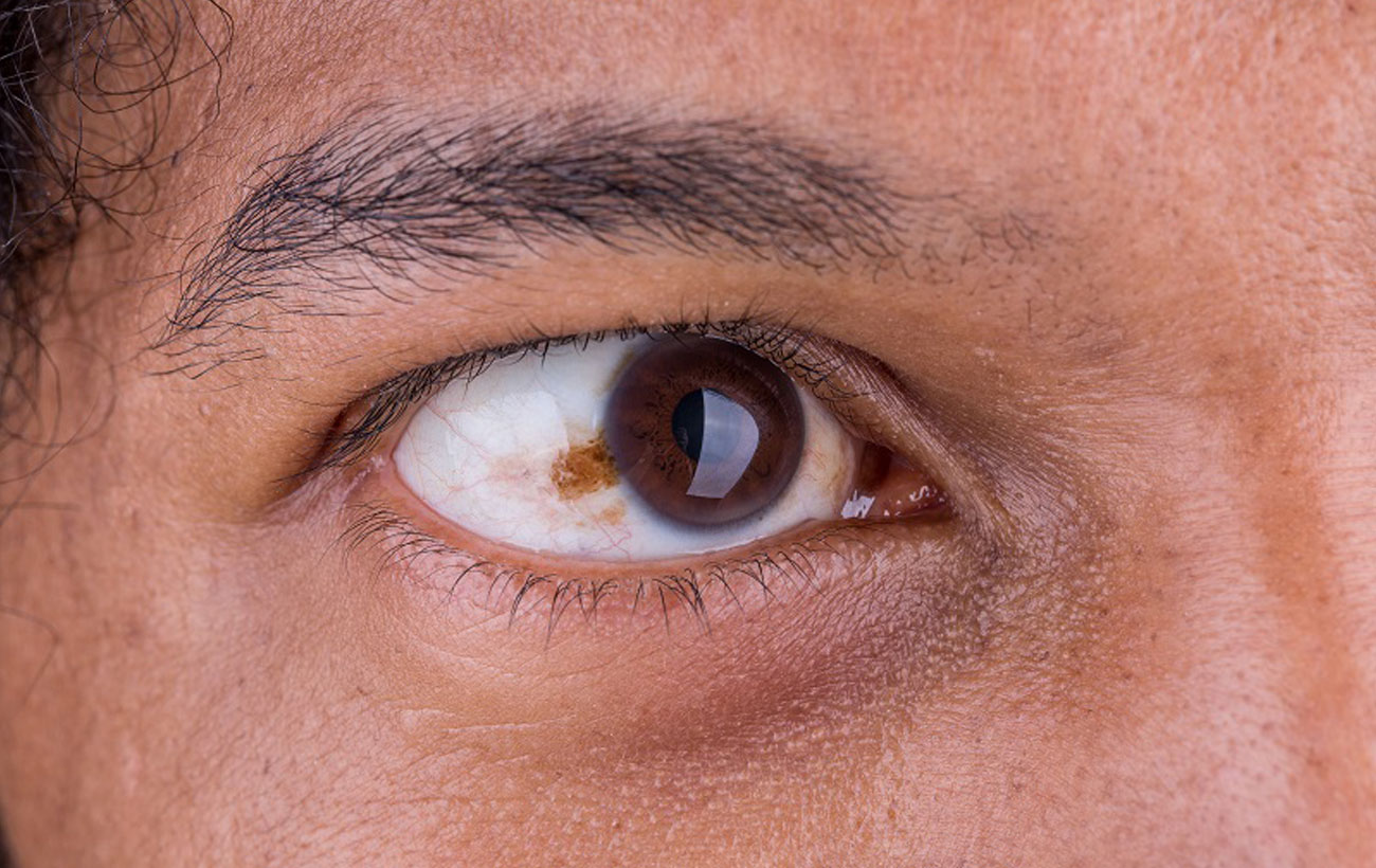
Malignant Melanoma
Malignant melanoma is a cancer that can affect the eyes as well as the skin,…

Retinoblastoma
Retinoblastoma is a rare, malignant tumor of the retina that occurs in one or both…

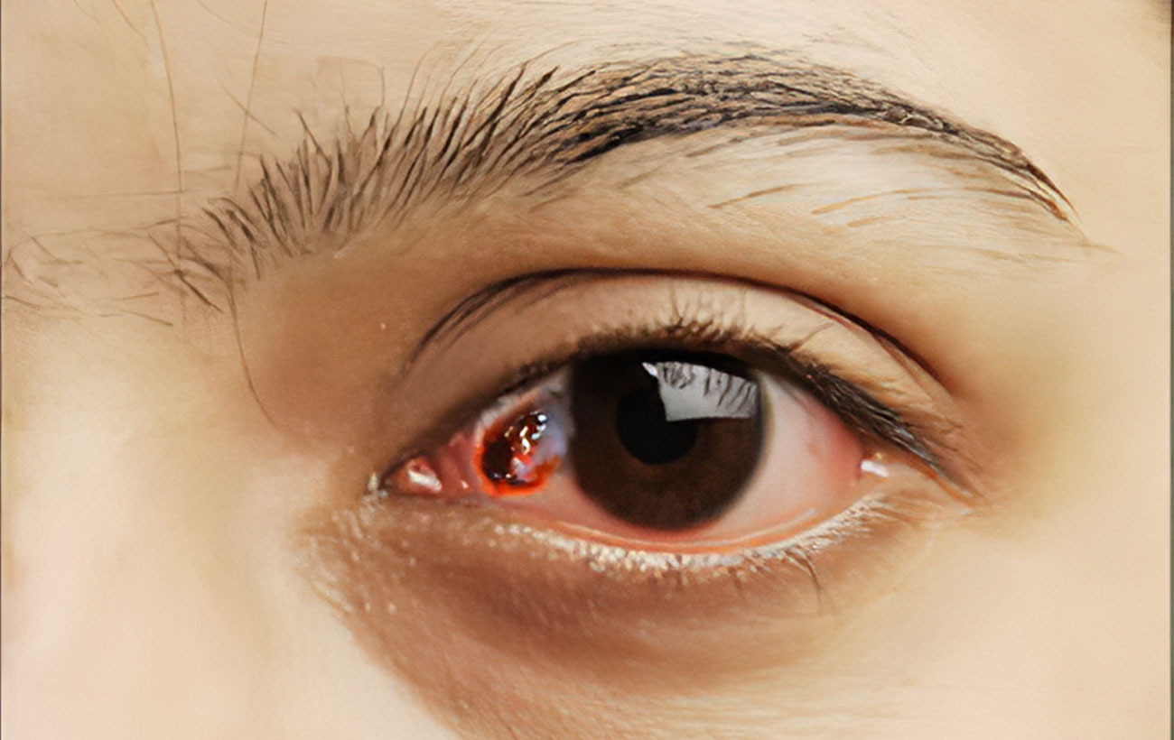
Subconjunctival Hemorrhage
A subconjunctival hemorrhage is leaking of blood from a small blood vessel in the eye…

Uveitis
Uveitis is inflammation of the uvea, which consists of the iris (the colored part of…

Watering Eye
A watering eye is an uncommon condition characterized by continuous tearing of the eye. Sometimes…

Corneal Ulcer
A corneal ulcer is an open sore or break in the surface of the cornea…

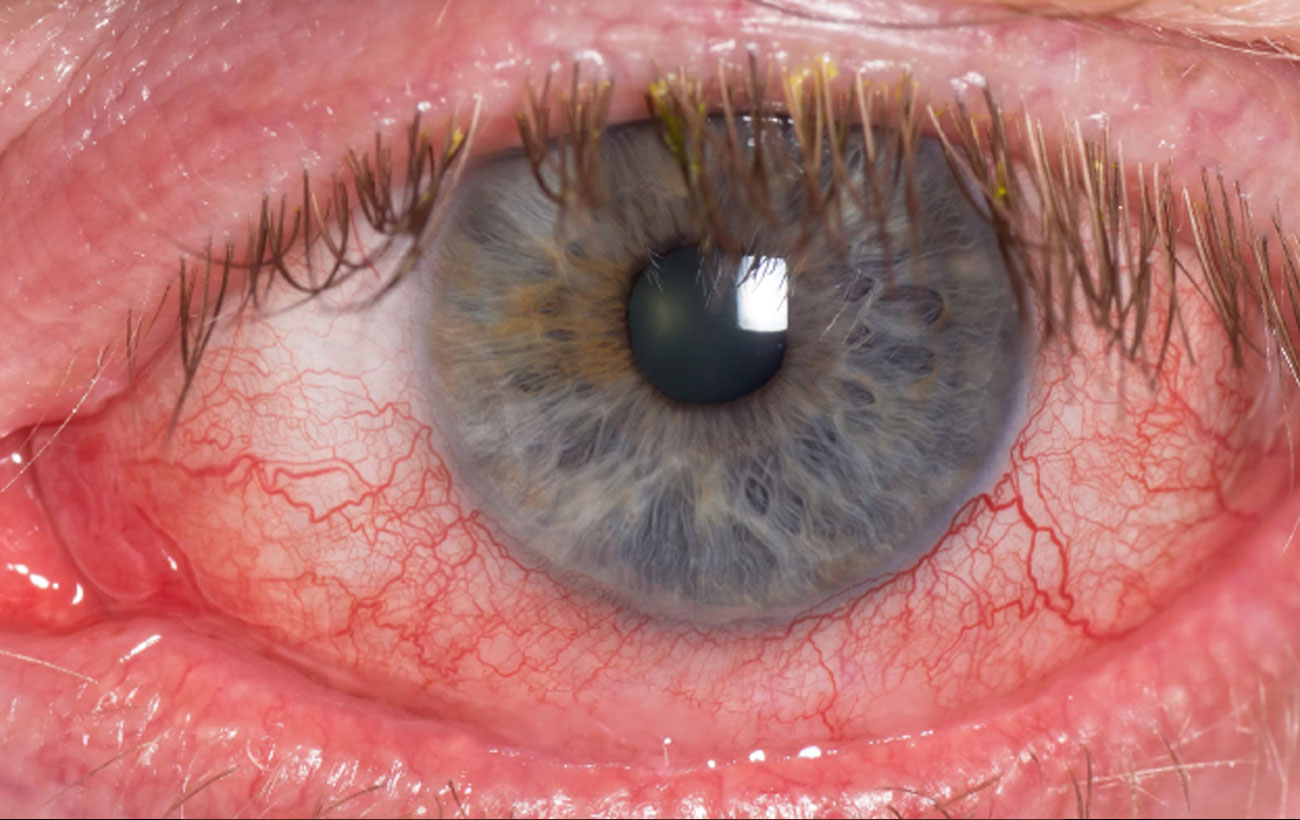
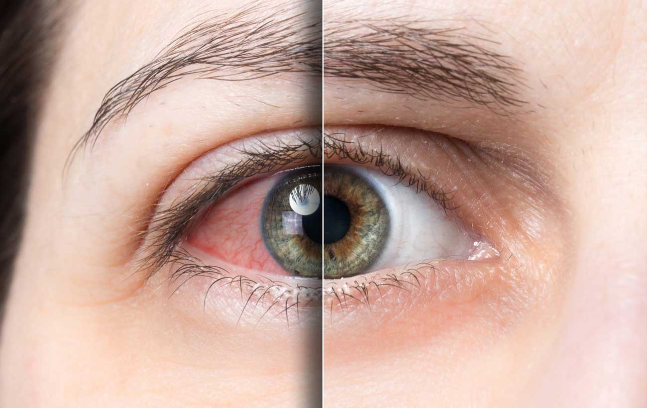
Conjunctivitis
Conjunctivitis (also called pinkeye) is inflammation of the conjunctiva, the transparent membrane that covers the…

Scleritis
Scleritis is inflammation of the sclera (the white of the eye). The condition is rare…
Dry eye syndrome
Dry eye is a condition that results from inadequate tear production. Although dry eye often…
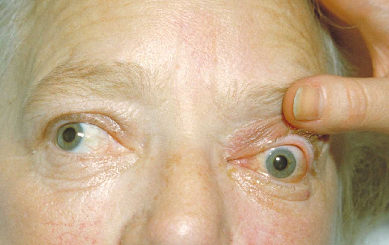
Exophthalmos
Exophthalmos is a condition in which one or both eyeballs bulge forward, exposing an abnormally…

Misaligned Eyes (Strabismus)
Normally aligned eyes move together and look in the same direction at the same time,…

Orbital Cellulitis
Orbital cellulitis is inflammation of the soft tissue of the orbit (the bony socket in…